Why We Work with Medical Radiologists

Approved by the American Board of Radiology.

Our radiologists come from some of the most prestigious institutions in the United States and are licensed in your state.

Going Beyond the Dentition.

Since CBCT scans can show anatomy outside of the dentition, there are still potential pathologies that a dentist, dental subspecialist, or even an Oral Maxillofacial Radiologist(OMR) might miss. A medical radiologist is better trained in the anatomy and in reviewing all of the other areas visible in a CBCT scan, meaning your patient might come in for a dental procedure and discover they have a potentially life-altering pathology such as head and neck cancer or carotid artery calcifications.

Head and neck cancer is the 7th most common type of cancer in the United States, resulting in 60,000 diagnosed cases and 11,000 deaths per year. The 5-year survival rate for head and neck cancers is 80% if caught early in stage 1 or 2. The survival rate drops to 40% if caught later at stage 3 or 4. Early detection makes all the difference in the outcome.

Asymmetrical carotid stenosis is the condition of a blockage in your carotid artery. According to the American Stroke Association, stroke ranks 5th on the list of conditions that cause death, and a leading cause of disability in the United States.

Because our medical radiologists are fully credentialed and licensed in the state where the CBCT scan was taken, they also help take the liability off of the dental practice.

Credentials and Training

We partner with Neuro Head and Neck radiologists who are approved by the American Board of Radiology and have completed:

  • 4 years of Medical School
  • 5 years of radiology residency
  • 1-2 years of a Neuroradiology + Craniofacial Imaging fellowship.

Your images will be interpreted by a full-credentialed expert, licensed in your state, who can take ownership of the entire scan.

Facilitated Referrals

If a critical finding is discovered, medical radiologists have the legal ability to create a diagnosis that insurance companies accept as a basis for patients to see a specialist.

With a medical referral, patients may not necessarily need to go through their primary physician to see a specialist, saving them critical time and money in getting the care they require.

Case Studies

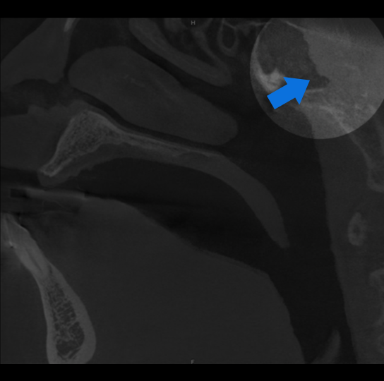
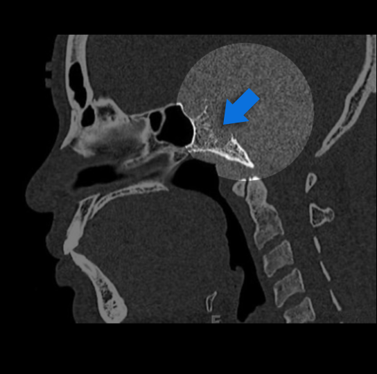
Below are a few examples of actual studies our radiologists have reviewed, all of which contained findings outside of the dentition. Drag the arrow across the image to view the specific location of the finding.
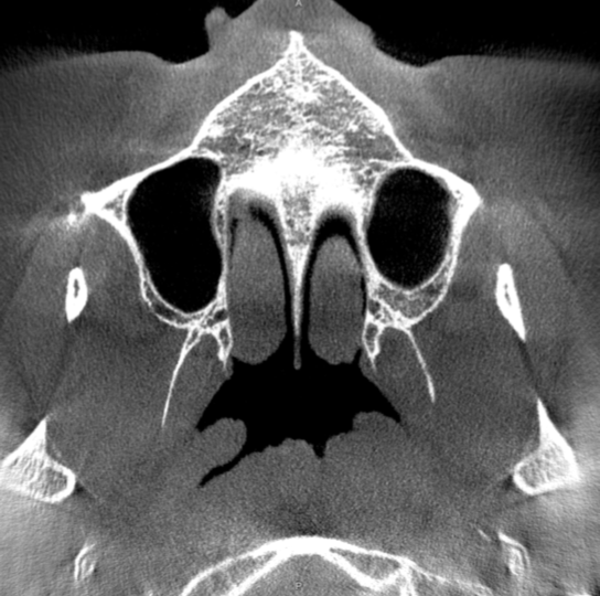
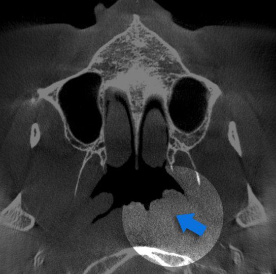
Study 1

Finding: Left Nasopharyngeal Mass.
Abnormal effacement of the left fossa of Rosenmuller, possibly representing a nonvisualized mass lesion. Direct visualization and ENT referral is recommended.
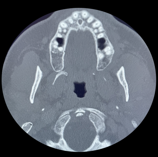
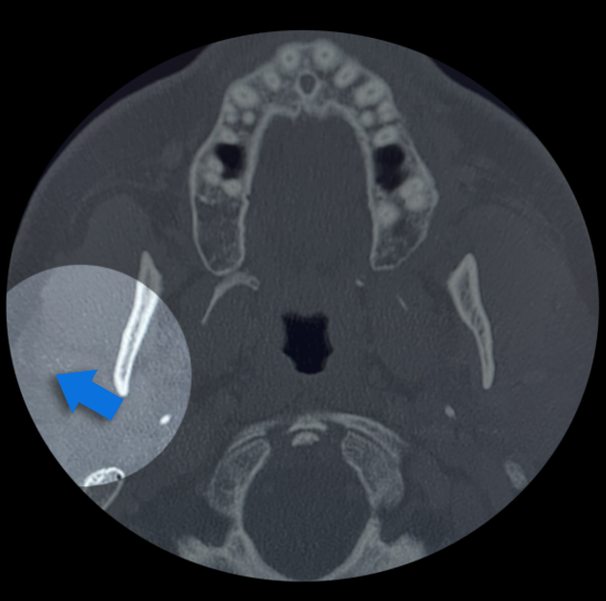
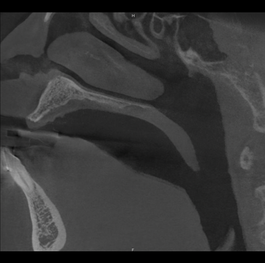
Study 2

Finding: Hypermetabolic mass in right parotid gland.
Suspicious subtle grouped calcifications in the right parotid gland. Using post processing technique to emphasize soft tissue, a subtle mass can be seen in the right parotid gland.
Study 3

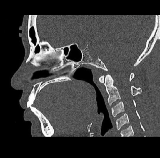
Finding: Congestion, airway “fullness”
Soft tissue mass in the right sphenoid sinus, erodes the posterior wall of sinus. Recommend MRI of the skull base to better evaluate.
Study 4

Finding: Lytic bone lesion of clivus
Suspicious lytic and erosive bone lesion involving the sphenoid and clivus. Recommend clinical referral and skull base MRI for more definitive evaluation. MRI follow up found an infiltrating soft tissue tumor, consistent with aggressive chordoma.

Photo of Ryan M. Walsh, D.D.S., M.S.

Although I feel I'm competent to interpret the scans, I just don't have the time to deeply interpret 80+ scans, other than for the specific tooth pathology I'm evaluating from an endodontic perspective. DentalRay gives me the peace of mind knowing I have a licensed radiologist reviewing the scans from a liability standpoint.

Ryan M. Walsh, D.D.S., M.S.
Advanced Endodontics of Texas

Elevate Your Standard of Care.

Create an account and upload your first scan today.